Unveiling Associations Between Retinal Microvascular Architecture and Phenotypes Across Thousands of Healthy Subjects
Michal Shapira, Smadar Shilo, Yeela Talmor-Barkan, Yaron Aviv, Yotam Reisner, Anastasia Godneva, Adina Weinberger, Alon Skaat, Anat Loewenstein, Eran Segal, Hagai Rossman
[paper]
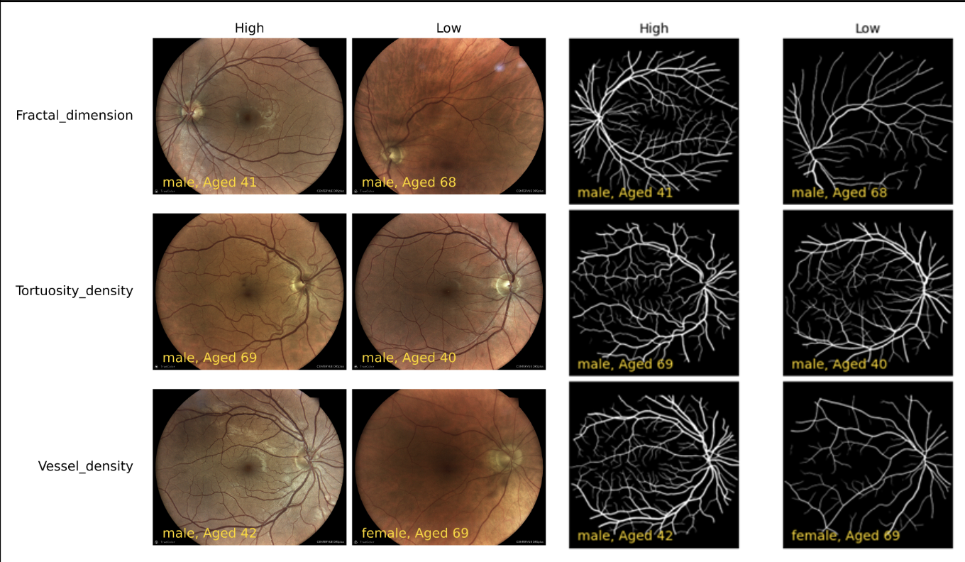
Fundus imaging has emerged as a powerful tool, allowing for high-resolution and non-invasive visualization of the fundus microvasculature. Recent advancements in artificial intelligence (AI) have enabled the quantification of fundus microvascular characteristics, paving the way for the development of predictive markers for systemic conditions. In this study, we characterize the fundus microvasculature among 8467 healthy individuals, aged 40-70 years. We employ a fully automated segmentation tool to calculate 12 distinct vessel measures, including tortuosity density, vessel density, average width, fractal dimension, distance tortuosity, and curvature tortuosity for both arteries and veins. We provide reference values for these measures by age and sex, and explore the associations between these fundus vessel measures and various clinical features. Our analysis revealed numerous significant correlations, including connections between fundus arterial measures and cardiovascular measurements, lipid profile, sleep apnea indices, glycemic profile, body composition, and smoking status. This comprehensive analysis of fundus microvasculature anatomy in a large healthy population contributes to the growing field of oculomics and highlights the potential of fundus imaging for earlier detection and monitoring of systemic diseases such as metabolic syndrome, cardiovascular diseases and sleep disorders.